Rock lobster larval diet. DNA barcoding from 1mm of tissue
2 minutes + 15 minutes (paper)
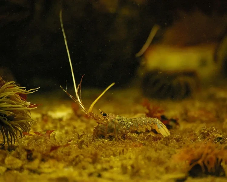
Western rock lobster larvae are largely transparent. Traditional microscopy cannot identify what is in their gut as the prey organisms are too soft and too similar in appearance to each other. This has made understanding larval feeding ecology, and therefore supporting lobster aquaculture, genuinely difficult.
A 2012 study published in PLOS ONE (O'Rorke et al.) used high-throughput amplicon sequencing of gut DNA from Panulirus cygnus phyllosomata to identify prey. Species identity of the larvae themselves was confirmed by sequencing the COI gene from a single 1mm pereiopod tip, extracted using the prepGEM kit in a 20 µl reaction volume.
The results revealed that the larvae were feeding predominantly on colonial radiolarians, thaliaceans, and other gelatinous zooplankton; groups that had never previously been identified in the larval gut using conventional methods. The work demonstrated that PCR-based gut content analysis from very small tissue samples is a viable approach to questions in marine ecology that microscopy simply cannot answer.
The extraction step: 1mm of leg tissue, 20 µl reaction, prepGEM; is a small but enabling component of a genuinely novel ecological finding.